Bony Tumour of Skullbase
Endoscopic SkullBase Surgery
Bony Tumour of Skullbase
Bony lesions of the skull base are a rare group of heterogeneous disorders. Clinical manifestation of these disorders often results from compression of the cranial nerves in the foramina at the skull base (optic nerve is most commonly affected), displacement of the eye and obstructing the nasal passage and sinuses.
Osteoma
Osteomas are relatively rare, slow-growing tumours. Most commonly they are found within the frontal and ethmoid sinuses. These tumours are slow growing and usually cause no symptoms. Small, asymptomatic lesions do not need surgery. Extension to the orbit and/or skull base is unusual. When osteomas expand, they give rise to symptoms by blocking the sinuses, like headache, and ocular symptoms, such as diplopia, exophthalmos and proptosis. On imaging, they tend to show a range of variable bone density, from very dense and sclerotic for ivory-type osteoma to less dense and less ossified for fibrous osteoma.
Surgery is the treatment of choice for symptomatic osteomas. However, the approach depends on the extension and the occurrence of complications. Traditional surgical approaches to the involved sinuses cut through the facial skin (external frontoethmoidectomy, lateral rhinotomy or osteoplastic flap technique). Endoscopic transnasal resection is ideal for tumours confined to the ethmoid and nasal cavity. The main advantages of the method are the minimal soft tissue dissection, the absence of facial bony disruption, and the avoidance of a facial incision. The magnification and the different angled view, which are possible with the use of endoscopes, may facilitate the removal of osteoma, with minimal morbidity. However, when osteomas are large and expanded in to the orbit and anterior cranial base, a combination of external and endoscopic technique are required, due to the limited access and visibility of endoscopy.
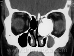
Osteoma – ethmoid; preoperative CT scan
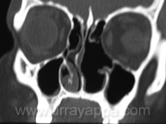
Osteoma – ethmoid; postoperative CT scan
Fibrous Dysplasia:
Fibrous dysplasia is an idiopathic, non-hereditary, progressive skeletal disorder in which the bone in localized areas being replaced by fibrous tissue resulting in expanded, softened, and fragile bone. It is established early in life and the activity continues until skeletal growth ceases. The most common bones affected by this disease are the skull and facial bones. Disease can present as monostotic fibrous dysplasia (affects only one bone), polyostotic fibrous dysplasia (involves multiple bones), or as McCune-Albright syndrome (polyostotic fibrous dysplasia with skin pigmentation and precocious puberty).
Fibrous dysplasia widens the medullary cavity, leaving the cortex intact. As a result, encroachment on the neurovascular foramina and paranasal sinus openings is often seen. CT is best for imaging and shows the inhomogeneous mixture of bone and fibrous tissue referred to as a “ground glass’’ appearance. A thin intact cortical rim is seen over the margins of the involved bones.
It is first detected in young children where it manifests as a swelling of the jaw. Involvement of the frontal and/or facial bones can eventually lead to deformation of facial features and skull shape, along with pain. When one or more bones progressively increase in size, they encroach on the cavities of the orbit, mouth the nose and sinuses. Abnormal protrusion of the eyeball (exophthalmos) may develop and eventually causes complete loss of sight due to compression of the optic nerve. There may also be interference of the nasal passage and with eating.
Asymptomatic lesions can be observed. Surgery is usually delayed until adolescence, however if the progression of the disease compromises neurological function, a decompressive procedure should be considered early in childhood to preserve normal function. Curative treatment for fibrous dysplasia involves complete removal of the affected bone. To preserve sight, surgery for optic nerve decompression is needed in cases where the fibrous dysplasia has deformed the optic canal. Some lesions are amenable to resection for a cure by a single procedure. More often, most lesions can be managed through staged procedures.
The endoscopic approach allows surgeons using the nose and nasal cavities to reach fibrous dysplasias that were once considered inoperable or hard to reach. It has the advantage of no facial scar or disfigurement to the patient, and a shorter recovery time.
Ossifying Fibroma
Ossifying fibroma is a neoplastic process affecting the bones of the skull. There is still confusion as to the distinctiveness of this disease, and these lesions have often been mislabeled as fibrous dysplasia, cementifying fibroma and fibrous osteoma. The distinct histological finding is the presence of osteoblasts and lamellar bone (fibrous dysplasia has woven bone with dysplastic bony spicules without osteoblasts). Head and neck lesions are most often in the mandible and maxilla, with rare occurrences reported in the paranasal and sphenoid bones and occasionally in the orbit. They tend to occur in the third and fourth decades, with the younger patients noted to have a more aggressive (faster growing) lesion.
Osteopetrosis
Osteopetrosis is a diffuse disease in which skeletal sclerosis is formed by thickening of both the cortical and the spongy bone and was known as marble bone disease. Most often patients present with deficits of cranial nerve (CN) II, VII and VIII. Blindness from optic nerve compression is almost exclusively associated with the juvenile autosomal recessive form of this disease.
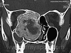
CT scan shows ossifying fibroma
The Heart Of Clinic

Dr. C. Rayappa MBBS, DLO, FRCS(Edin)
SENIOR CONSULTANT
+91 44 3315 1105
Dr. C. Rayappa graduated from Madras Medical College, Chennai, India in 1982. He completed his post graduation in Otolaryngology (ENT)