Cancer of the Ear
Head and Neck Tumour Surgery
Cancer of the Ear
Anatomy of the ear
The ear has three parts – outer ear, middle ear and inner ear (Fig.1). The outer ear is made of external ear (pinna) and external ear canal. Ear drum (tympanic membrane) is at the medial end of ear canal and it separates the outer ear from middle ear. The middle ear lies medial to the ear drum. Eustachian tube connects the middle ear to the nasopharynx. Medial to the middle ear lies the inner ear concerned with hearing and balance. The inner ear has three parts viz. cochlea, labyrinth and semicircular canals. From the inner ear nerve goes to the brain. The inner ear, middle ear and part of the outer ear canal are housed in the temporal bone. The nerve that controls the movement of face (facial nerve) runs above the inner ear and through the middle ear. The internal carotid artery and internal jugular vein & sigmoid sinus are closely related to the temporal bone.

Fig.1. EAC-External auditory Canal, TM-Tympanic membrane, JB-Jugular bulb, ICA-Internal carotid artery, IJV-internal jugular vein, Lab-Labyrinth, SCC-semi circular canal, ME-Middle ear.
What type of cancers occur in the ear?
Cancer of the ear is rare. It can occur in any part of the ear (external ear or ear canal, middle ear or inner ear). Those that develop inside the inner ear are rare. Most of the cancers are squamous cell carcinomas. Other types include basal cell cancer, melanoma, adenoid cystic carcinoma and adenocarcinoma.
What causes it?
These cancers are usually found in individuals who have neglected long-term drainage and infection in the mastoid or middle ear. The exact relationship of the infection to the formation of the squamous cell cancer is unclear.
What symptoms does it cause?
Squamous cell cancer of the middle ear is often quite advanced before a correct diagnosis is made. Pain is a significant feature of squamous cell cancer of the middle ear and mastoid. Intermittent bleeding and drainage for long periods of time are also usual. Hearing loss is significant. Some people may also have swollen lymph nodes in their neck. Occasionally people cannot move their face on the side of the affected ear.
How is it diagnosed?
Generally an early stage cancer is small and just within the area it started in. Later it grows into the surrounding structures and much later it spreads to another part of the body. Diagnosis depends upon a biopsy of the tissue. Surgeon will take a small amount of tissue from the abnormal area of the ear and to examine it under a microscope. If the biopsy shows a cancer, surgeon will do CT scan and MRI scan to know the extent of disease to decide which treatment is needed. PET scan is done to rule out spread of tumour to other parts of the body.
How is it treated?
The main treatments for these cancers are surgery and radiotherapy. Depending on the stage of the cancer, patient may also have chemotherapy. The treatment depends on type of cancer, size of tumour, which part of the ear is involved, spread to adjacent structures like parotid salivary gland, dura, brain as well as general health of the patient.
Surgery
The type of surgery needed depends on the area of ear affected, and whether it has spread into nearby structures, such as the bone, dura, parotid etc. Surgeon will remove the tumour together with an area of tissue surrounding it that is completely free of cancer cells. Doing this helps to lower the risk of the cancer coming back.
- Surgery may involve having some or all of the following removed.
- The ear canal
- Part or all of the temporal bone
- The middle ear
- The inner ear
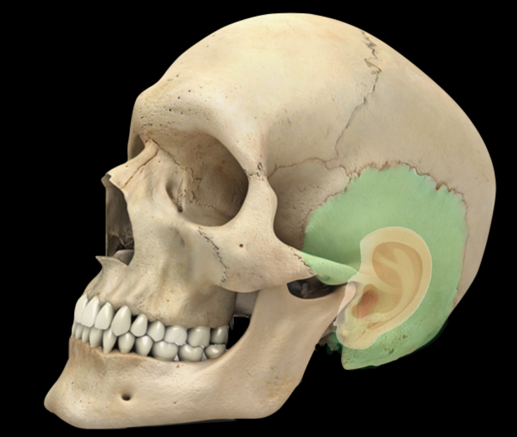
Fig.2. Temporal bone is shaded green at the side of the skull, medial to the pinna.
The temporal bone is the bone at the side of the skull, adjacent to the external ear (Fig.2). The operations to remove the temporal bone tumour may range from a mastoidectomy to total temporal bone resection.

They may also need to remove the lymph nodes nearby in the neck and the salivary gland on that side of the head. Rarely, surgeon may need to remove the facial nerve, which runs through the temporal bone. In that case, nerve grafting is done to regain the facial movement.
If the middle and inner ear were removed the patient will be able to hear on that side. Balance may be affected and the patient may feel dizzy temporarily. Sometimes it is possible for the surgeon to rebuild (reconstruct) some of the ear so that hearing can be preserved. With this operation, appearance won’t change for most people. Scar line is likely to be behind the ear and will not be visible .
Radiotherapy
Radiotherapy uses high-energy rays to treat cancer. Radiotherapy may be taken as main treatment in early lesions (when there is no bone erosion) or along with surgery if there is bone erosion or if the surgeon hasn’t been able to remove a clear margin of tissue from around the tumour. Then radiotherapy can lower the risk of the cancer coming back.
Chemotherapy
Chemotherapy uses anti cancer (cytotoxic) drugs to destroy cancer cells. Chemotherapy on its own won’t cure cancer of the ear but doctors may use it to relieve symptoms if your cancer comes back or you can’t have other treatments.
The Heart Of Clinic

Dr. C. Rayappa MBBS, DLO, FRCS(Edin)
SENIOR CONSULTANT
+91 44 3315 1105
Dr. C. Rayappa graduated from Madras Medical College, Chennai, India in 1982. He completed his post graduation in Otolaryngology (ENT)
For Doctors
Need more information?
Reach us through mail or phone
The Heart Of Clinic
Dr. C. Rayappa MBBS, DLO, FRCS(Edin)
SENIOR CONSULTANT
+91 44 3315 1105
Dr. C. Rayappa graduated from Madras Medical College, Chennai, India in 1982. He completed his post graduation in Otolaryngology (ENT)