Open Skull Base Surgery
Anterior skull base surgery: open
Where is the skull base?
The skull base (Fig.1) is the bony surface under the brain.
There are number of structures lie below the skull base viz. nose & sinuses, eyes, ears etc. Major blood vessels taking blood to and from brain, nerves of the eye, ear and those controlling facial movement, swallowing & speech and spinal cord passes through number of natural foramina in the skull base.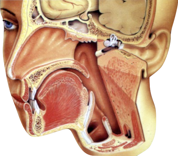
Fig.1
Which Types of Diseases Are Treated With Skull Base Surgery?
The skull base tumours or tumour-like conditions may arise from the skull base bone itself. Or it may arise from the brain, covering of the brain (dura) or from the nerves. Sometimes these may arise from extra cranial structures like nose & sinuses, eye or ear and encroach on the skull base.
A variety of benign (non-cancer) tumours occur at the skull base. These include acoustic neuroma, meningioma, schwannoma, glomus jugulare, epidermoid, pituitary tumours, and many others. In uncontrolled diabetics, infection of skull base bone (osteomyelitis) takes place.
Malignant (cancer) tumours managed with skull base surgery include squamous cell carcinoma, esthesioneuroblastoma, chondrosarcoma, and chordoma.
What type of symptoms do patients with skull base tumours have?
The skull base tumours are rare and usually cause few symptoms until they grow to a size where they begin to affect neurologic function. These symptoms may manifest as loss of sight, double vision, nosebleeds, facial pain or twitching, hearing loss, dizziness, hoarseness, tongue weakness or hormonal disturbances.
How is a skull base tumour diagnosed?
These symptoms most often come on gradually and are diagnosed by an ENT specialist and/or neurosurgeon using a CT scan and MRI. Specialized tests such as hearing tests, visual field testing, or arterial studies may also be performed. For very vascular tumours, an angiogram with embolization may be performed prior to surgical resection in order to plug the arteries that feed the tumour. This reduces blood loss during surgery.
What Technologies Are Used During Skull Base Surgery?
Skull base is a complex anatomical region. To improve the safety of surgery, surgeons use number of gadgets.
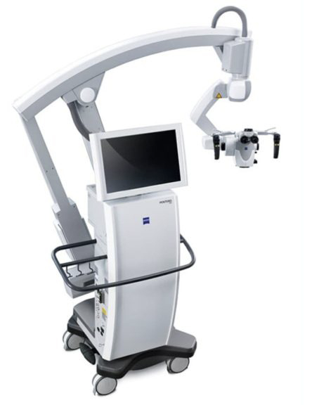
Fig.2. Operating microscope
Surgeons use high-power microscope (Fig.2) to magnify the operation site for proper visualization of vital structures. This helps in meticulous removal of tumour without injuring the adjacent vital structures.
Image Guidance During Surgery
Many times, surgery is guided by computer software that incorporates MRI and CT scans of the tumour. Using a probe referenced to the patient on the operating table, the surgeon can pinpoint the location of the tumour and verify exactly where each vital structure is in relation to the tumour (Fig.3). It is also used to determine the best possible surgical route to the tumour, which may be located deep within the skull base.
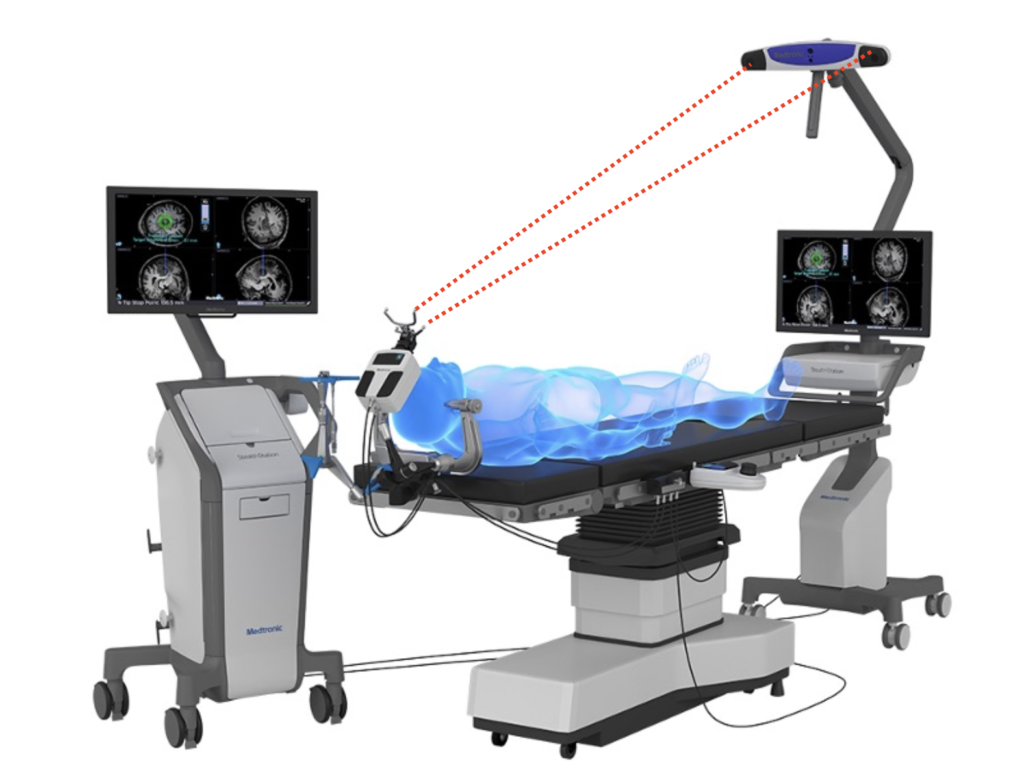
Fig.3. Neuronavigation

Fig.4.
Neurophysiological Monitoring
Even with detailed knowledge of anatomy and surgical skill, motor nerves can sometimes be difficult to identify during surgery due to disease, a previous operation, or normal anatomical variations. For example, during skullbase surgery, the facial nerve is commonly exposed and at risk for injury. Since this nerve controls all movements and expressions of the face, damaging this nerve can have devastating physical and emotional results. Patients can suffer temporary or permanent damage if a nerve is irritated or injured. Intraoperative nerve monitors (Fig.4) enable surgeons to identify, confirm, and monitor motor nerve function during a variety of surgical procedures to help reduce the potential risk of nerve damage.
How is this surgery performed?
Skull base surgery is performed by a multidisciplinary team consisting ENT-Head & Neck Surgeon, Neurosurgeon and Plastic surgeon to provide the best possible outcome.
Tumours of the skull base are underneath the brain and it can be difficult to reach the tumour during surgery. In traditional techniques significant force is used to retract the patient’s brain out of the way. This may lead to unwanted injury to otherwise normal brain tissue.The skull base team uses innovative surgical techniques which reduces the retraction of normal brain, protects uninvolved neurovascular structures and does not compromise tumour removal.
Some tumours, usually those located in the central part of the skull base (in close proximity to nose and sinuses), may be removed by surgery performed through the nostrils without opening the skull, known as ‘endoscopic skull base surgery’ (detailed information about this minimally invasive surgery of skull base is given in Endoscopic Skull Base Surgery section). Sometimes, due to a tumour’s location or size, it may need to be removed through open surgery.
Skull base procedures, for example, may be designed to traverse the bone containing the ear (trans-temporal), low on the temple beneath the brain (middle fossa approach), around the eye (trans-orbital/orbital-zygomatic/craniofacial approaches), through the nose or paranasal sinuses (trans-sphenoidal/trans-facial approaches), or from the neck (trans-cervical).
Success rates of skull base surgery have continued to improve. This is because of the evolution of modern approaches designed to reduce the requirement of brain retraction and sacrifice of normal structures in order to get to the tumour.
Our surgical team is most experienced in both open and minimally invasive skull base surgical techniques. They develop the most effective surgical plan for the patient, depending on the location of the tumour.
If a tumour can’t be completely removed, additional treatments—such as radiation therapy or chemotherapy may be needed.
What Are the Treatment Options?
Not all skull base conditions require surgery. Most skull base tumours are benign, or non-cancer . However, they can put pressure on major nerves and cause serious symptoms when they grow. Minimally invasive surgery to remove these types of tumours focuses on preserving nerve function.
Some skull base tumours are malignant, or harmful, and must be removed. They may require chemotherapy and/or radiation therapy before or after surgery.
What Questions Should I Ask My Doctor?
- What type of skull base condition/tumour do I have?
- What symptoms should I expect if this continues to grow?
- Are there multiple treatment options, and what are the risks and benefits of each?
- Which medical/surgical specialists will be involved in my care?
- Is the surgeon having enough experience in open as well as minimally invasive surgery?
- What is my expected long-term outcome with this condition?