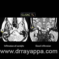
Fig.1 MRI shows carcinoma of EAC infiltrating the mandibular condyle and middle fossa dura
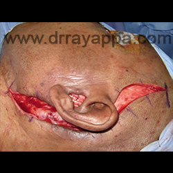
Fig.2 Postaural incision and EAC incision.
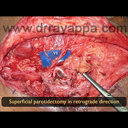
Fig.3 Retrograde parotidectomy done. Parotid wasn’t seperated from EAC.
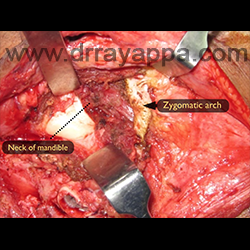
Fig.4 Exposed neck of mandible is the site of osteotomy ( dotted line) for condylectomy.
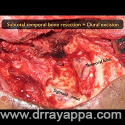
Fig.5 Subtotal temporal bone resection had been done. Middle fossa dura that was infiltrated by the tumour was excised. Picture shows exposed temporal lobe of brain, ICA and sigmoid sinus.
Fig.6 Dural defect was reconstructed with fascia lata. Facial nerve was anastamosed to hypoglossal nerve. Cavity was filled with free fat graft.
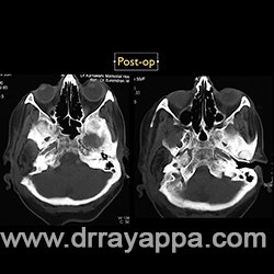
Fig.7 Post-op (3 yrs) shows no recurrence.
The Heart Of Clinic
Dr. C. Rayappa MBBS, DLO, FRCS(Edin)
SENIOR CONSULTANT
+91 44 3315 1105
Dr. C. Rayappa graduated from Madras Medical College, Chennai, India in 1982. He completed his post graduation in Otolaryngology (ENT)