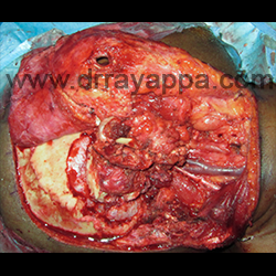
Fig.3 Craniectomy done to expose middle and posterior fossa dura. Mandible is divided and TMJ disarticulated.
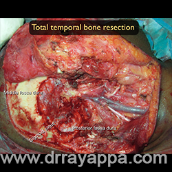
Fig.4 Total temporal bone resection done. Picture shows the miidle & posterior fossa dura , sigmoid sinus, lower cranial nerves and IJV.
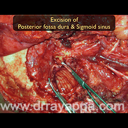
Fig.5 Since the posterior fossa dura was infiltrated by the tumour, it was excised along with sigmoid sinus. Cerebellum is seen through the dural defect.
The Heart Of Clinic
Dr. C. Rayappa MBBS, DLO, FRCS(Edin)
SENIOR CONSULTANT
+91 44 3315 1105
Dr. C. Rayappa graduated from Madras Medical College, Chennai, India in 1982. He completed his post graduation in Otolaryngology (ENT)