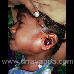
Fig.1 2 year old male. Swelling infront of left year with a fleshy mass coming out of ear canal and blood stained discharge for 3 months.
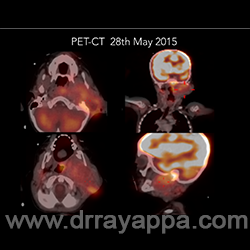
Fig.2 PET – CT shows hypermetabolic mass involving the ear and infratemporal fossa.
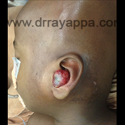
Fig.3 Residual tumour after chemotherapy.
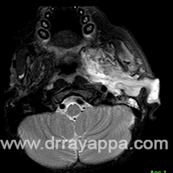
Fig.4 MRI shows residual tumour in infratemporal fossa, middle ear and external ear.
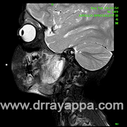
Fig.5 MRI shows residual tumour in infratemporal fossa.
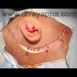
Fig.6 Postaural incision from temporal region to submandibular region. Another circumferential incision ar the lateral end of EAC.
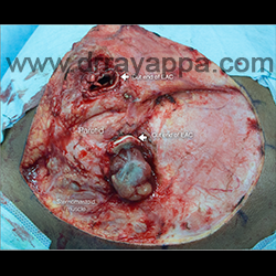
Fig.7 Skin flap is raised after dividing the lateral end of external auditory canal. Picture shows the parotid gland, sternomastoid muscle, temporais muscle and cut ends of EAC.
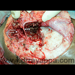
Fig.8 Sternomastoid and temporalis muscle were dissected off bony attachment and mastoid tip. Superficial lobe of parotid gland is elevated
off the facial nerve trunk and its branches
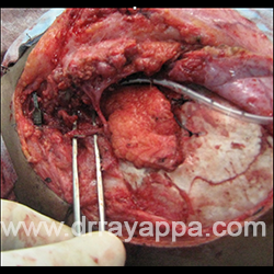
Fig.9 Lateral temporal bone resection & condylectomy had been done. Facial nerve and all its branches were preserved.
Fig.10 Cavity is filled with anterolateral thigh microvascular flap.
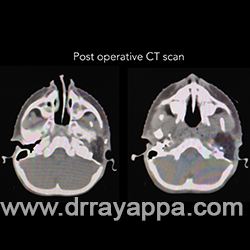
Fig.11 Post-op CT scan shows no residual recurrent tumour.
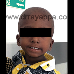
Fig.12 6 months poat-op. Good cosmesis and facial nerve
The Heart Of Clinic
Dr. C. Rayappa MBBS, DLO, FRCS(Edin)
SENIOR CONSULTANT
+91 44 3315 1105
Dr. C. Rayappa graduated from Madras Medical College, Chennai, India in 1982. He completed his post graduation in Otolaryngology (ENT)