drrayappa.com

Carcinoma of External Auditory Canal (T1)
Lateral Temporal Bone Resection

Carcinoma of External Auditory Canal (T1) Lateral Temporal Bone Resection

Fig.1
Fig.2 Lateral temporal bone resection – for T1 tumours limited to EAC
Fig.3 CT scan shows tumour (yellow arrow) confined to bony EAC. No bony erosion. No extension into middle ear
Fig.4 Circle – incision at the lateral end of EAC. Dotted line – post aural incision
Fig.5 Extended cortical mastoidectomy & extended posterior tympanotomy done
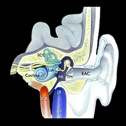
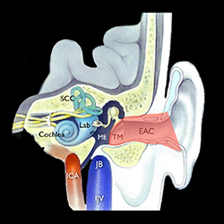
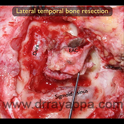
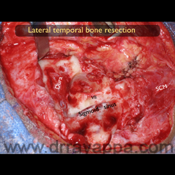
Fig.6 After cutting through the tympanic bone and disarticulating the ossicles, the entire EAC is removed along with tympanic membrane. Picture shows exposed middle fossa dura, sigmoid sinus and mastoid segment of facial nerve. SCM – sternomastoid

The Heart Of Clinic

Dr. Rayappa

Dr. C. Rayappa MBBS, DLO, FRCS(Edin)

SENIOR CONSULTANT

+91 44 3315 1105

Dr. C. Rayappa graduated from Madras Medical College, Chennai, India in 1982. He completed his post graduation in Otolaryngology (ENT)

The Heart Of Clinic

Dr. Rayappa

Dr. C. Rayappa MBBS, DLO, FRCS(Edin)

SENIOR CONSULTANT

+91 44 3315 1105

Dr. C. Rayappa graduated from Madras Medical College, Chennai, India in 1982. He completed his post graduation in Otolaryngology (ENT)