Skip to main content
drrayappa.com
Home
Profile
Specialities
Menu
Head and Neck Tumour Surgery
Open Skull Base Surgery
Lateral Skull Base Surgery
Endoscopic Pituitary Surgery
Endoscopic SkullBase Surgery
Skull Base Surgery In Children
Surgical Video
Menu
Head and Neck Tumour Surgery
Open Skull Base Surgery
Lateral Skull Base Surgery
Endoscopic Pituitary Surgery
Endoscopic SkullBase Surgery
Skull Base Surgery in Children
International Patients
Testimonial
Newsletter
Contact
Quick Links
Acromegaly, Gigantism
Angiofibroma
Anterior Clinoid Mucocoele
Brain Fluid Leak
Carotid Body Tumour
Cheek Cancer
Clival Chordoma
Craniofacial Resection
Craniofacial Resection
CSF Rhinorrhoea
Cushing’s Disease
Ear Cancer
Endoscopic Craniofacial resection
Endoscopic Nasopharyngectomy
Endoscopic Pituitary Surgery
Endoscopic Skullbase Surgery
Esthesioneuroblastoma
Ethmoid Cancer
Facial Nerve Tumour
Facial Translocation Approach
Far Lateral Approach Approach
Gum / Gingival Cancer
Glomus Jugulare
Glomus Tympanicum
Glomus Vagale
Hypopharynx Cancer
Hemithyroidectomy
Larynx Cancer
Laser Cordectomy
Lower Jaw Cancer
Maxillary Sinus Cancer
Maxillectomy
Meningocoele
Midline Labiomandibuloglossotomy
Mouth Cancer
Nasopharynx Cancer
Nasopharyngectomy
Odontoidectomy
Optic Nerve Decompression
Oral Cancer
Osteogenic Sarcoma
Parotidectomy
Parotid Tumour
Partial Laryngectomy
Pediateric Skullbase Surgery
Petrous Apex Cholesterol Granuloma
Petrous Apex Cyst
Pituitary Tumour
Postcricoid Cancer
Prolactinoma
Pyriform Sinus Cancer
Radioactive Lodine Treatment
Rhabdomyosarcoma
Schwannoma of Infratemporal Fossa
Sinus Cancer
Skullbase surgery in children
Speech After Laryngectomy
Stomal Recurrence
Temporal Bone Resection
Teratoma of Sella
Throat Cancer
Thyroid Cancer
Tongue Cancer
Tonsil Cancer
Total Laryngectomy
Total Thyroidectomy
Transcondylar Approach
Trans Oral Robotic Surgery TORS
Trans Oral Ultrasonic Surgery TOUSS
Vocal Cord Cancer
Upper Jaw Cancer
X
Chordoma - Pre & Post-op MRI
Chordoma - Pre & Post-op MRI
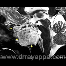
Fig.1
Pre-op sagittal MRI shows chordoma (yellow arrows) which has destroyed the sphenoid sinus and part of clivus.
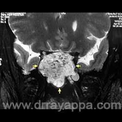
Fig.2
Pre-op coronal MRI shows chordoma (yellow arrows) which has destroyed the sphenoid sinus and part of clivus.
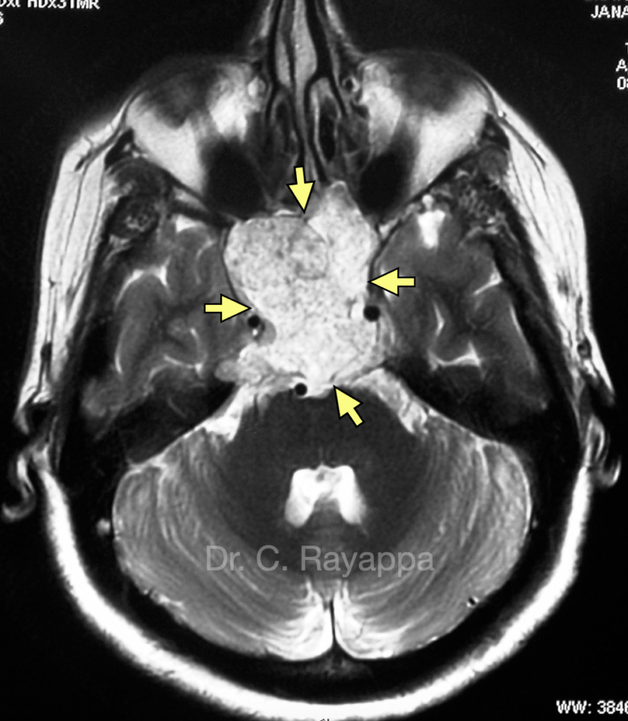
Fig.3
Pre-op axial MRI shows chordoma (yellow arrows) which has destroyed the sphenoid sinus and part of clivus.
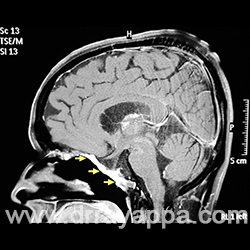
Fig.4
Post-op MRI shows complete excision of tumour. Yellow arrows are pointing at the Hadad’s nasoseptal flap used for reconstruction.
The Heart Of Clinic
Dr. C. Rayappa MBBS, DLO, FRCS(Edin)
SENIOR CONSULTANT
+91 44 3315 1105
Dr. C. Rayappa graduated from Madras Medical College, Chennai, India in 1982. He completed his post graduation in Otolaryngology (ENT)
Learn More
For Doctors
Chordoma with intradural extension + VIDEO
Chordoma in 11yrs old boy
Chordoma - Pre & Post-op MRI
Need more information?
Reach us through mail or phone
The Heart Of Clinic
Dr. C. Rayappa MBBS, DLO, FRCS(Edin)
SENIOR CONSULTANT
+91 44 3315 1105
Dr. C. Rayappa graduated from Madras Medical College, Chennai, India in 1982. He completed his post graduation in Otolaryngology (ENT)
Learn More
pdf download popup
Please enable JavaScript in your browser to complete this form.
Name
*
First
Last
Email
*
Message Comment Name
Comment or Message
Submit
×