Congenital Sphenoid Meningoencephalocoele in 3 yr old. Pre & Post op MRI
Congenital Sphenoid Meningoencephalocoele in 3 yr old. Pre & Post op MRI
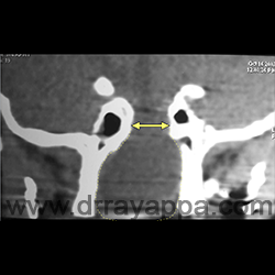
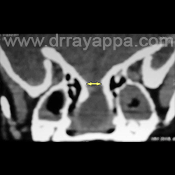
Fig.1 2 yr old boy. Complete nasal obstruction, watery nasal discharge and recurrent meningitis. CT shows a large bony defect in the sphenoid roof with soft tissue mass filling the nasal cavity and nasopharynx.
Fig.2 CT shows a large bony defect in the sphenoid roof with soft tissue mass filling the nasal cavity.
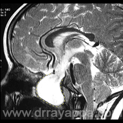
Fig.3 MRI shows a large meningoencephalocoele filling the nasal cavity and nasopharynx.
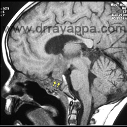
Fig.4 1 yr post endoscopic repair. MRI shows good nasal airway, soft tissue covering the area of skull base repair. Yellow arrows pointing at a black line formed by the bone used to repair the skull base defect.
The Heart Of Clinic
Dr. C. Rayappa MBBS, DLO, FRCS(Edin)
SENIOR CONSULTANT
+91 44 3315 1105
Dr. C. Rayappa graduated from Madras Medical College, Chennai, India in 1982. He completed his post graduation in Otolaryngology (ENT)