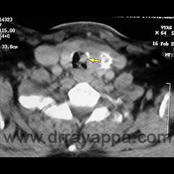
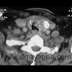
Fig.2 CT scan shows tumour involving left lobe of thyroid and inside tracheal lumen (yellow arrow).
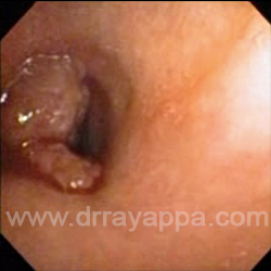
Fig.3 Endoscopy picture of tumour inside trachea.
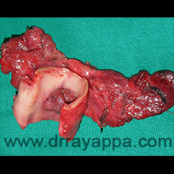
Fig.4 Picture shows operated specimen with entire thyroid gland, resected tracheal segment and the tumour within tracheal lumen.
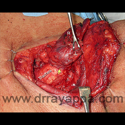
Fig.5 Both the lobes of thyroid are dissected leaving it attached to the left side of trachea where it is infiltrated. Yellow arrow – site of tracheal invasion. ‘R’ – right lobe of thyroid. ‘L’ – left lobe of thyroid.
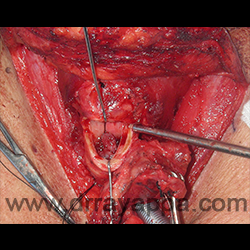
Fig.6 Trachea is cut above and below the tumour and the segment of trachea is resected along with the thyroid. Picture shows thyroid gland, upper cut in trachea and tumour within the tracheal lumen (yellow arrow).
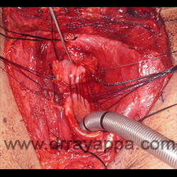
Fig.7 Posterior wall of trachea is being sutured together.
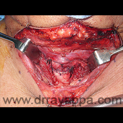
Fig.8 Anterior wall of trachea is sutured together.
The Heart Of Clinic
Dr. C. Rayappa MBBS, DLO, FRCS(Edin)
SENIOR CONSULTANT
+91 44 3315 1105
Dr. C. Rayappa graduated from Madras Medical College, Chennai, India in 1982. He completed his post graduation in Otolaryngology (ENT)