Meningioma Of Lower Clivus Surrounding Vertebral Artery
Meningioma Of Lower Clivus Surrounding Vertebral Artery
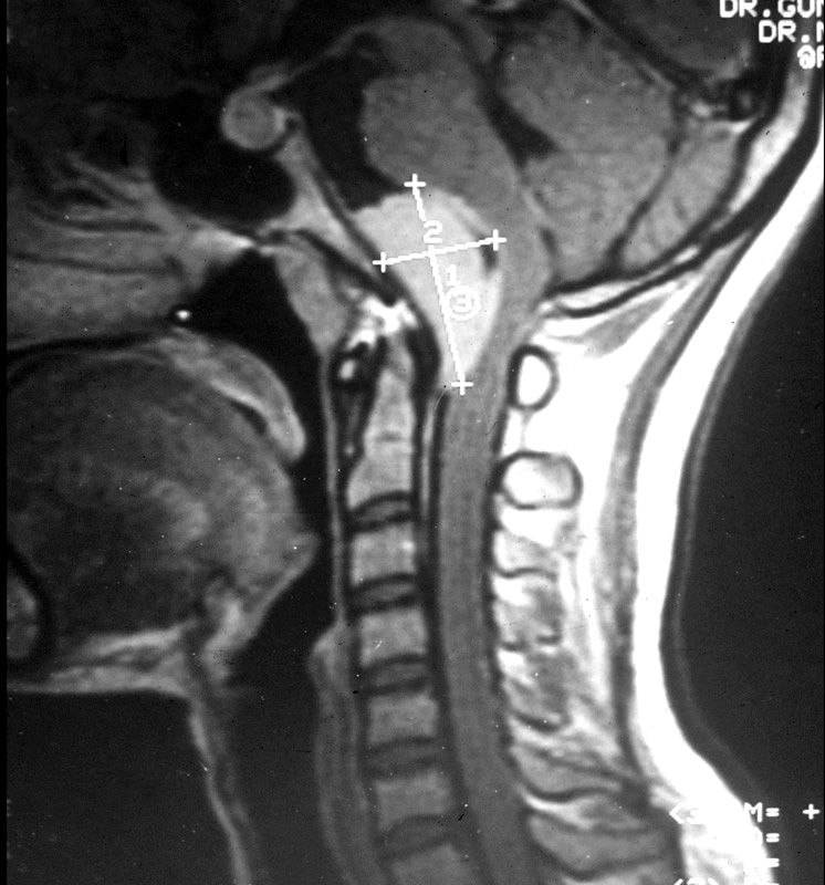
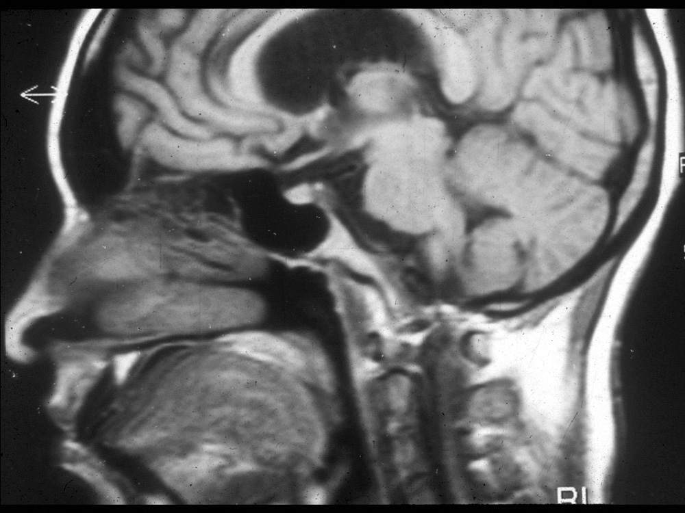
Fig.1 Sagittal MRI shows meningioma behind the lower clivus and atlas pressing the brain stem.
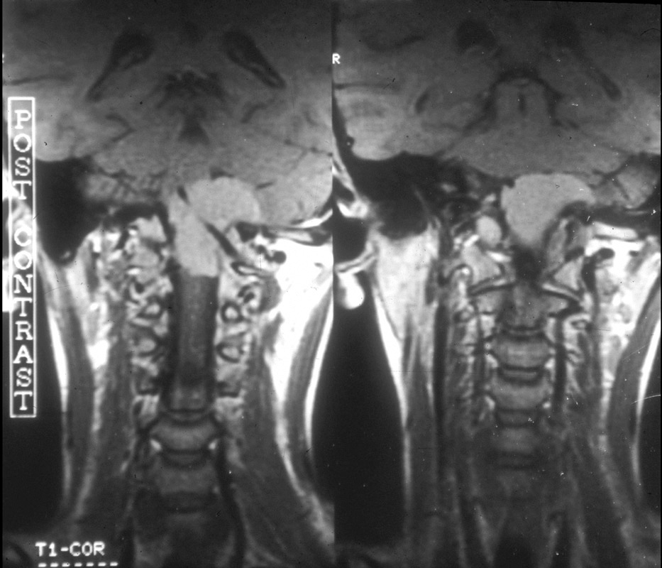
Fig.2 Coronal MRI shows the left vertebral artery going through the tumour.
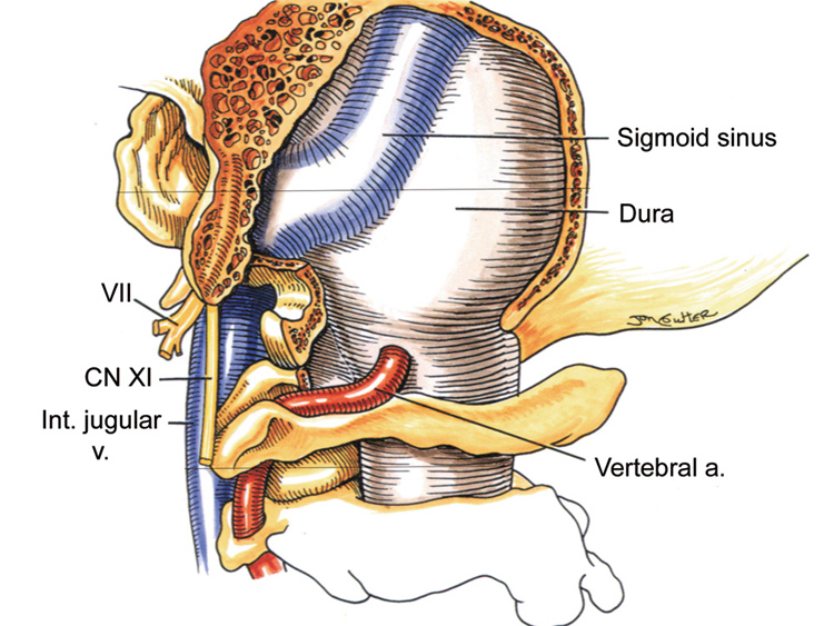
Fig.3 In far lateral approach, retrosigmoid craniectomy extended inferiorly upto foramen magnum, Vertebral artery is rerouted posteriorly after opening foramen transversorium in atlas. One third of occipital condyle can be resected
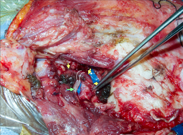
Fig.4 Oblique muscles were removed. Tip of C1 transverse process is removed to open foramen transversorium (two white lines). Yellow arrows points at the the vertebral artery surrounded by venous plexus. Green arrow – posterior arch of atlas.
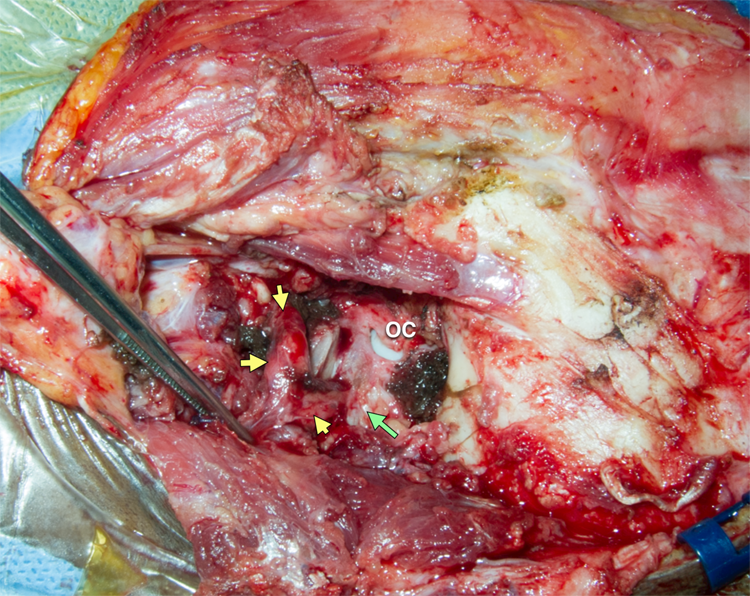
Fig.5 Posteriorly rerouted vertebral artery (yellow arrows) from C2 to the dural entry point. OC – occipital condyle. Green arrow – foramen magnum.
Fig.6 Posteriorly rerouted vertebral artery (yellow arrows) from C2 to the dural entry point. OC – occipital condyle. Green arrow – foramen magnum.
Fig.7 Post-op MRI shows complete resection of tumour.
The Heart Of Clinic
Dr. C. Rayappa MBBS, DLO, FRCS(Edin)
SENIOR CONSULTANT
+91 44 3315 1105
Dr. C. Rayappa graduated from Madras Medical College, Chennai, India in 1982. He completed his post graduation in Otolaryngology (ENT)