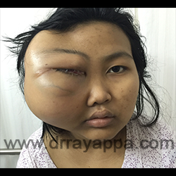
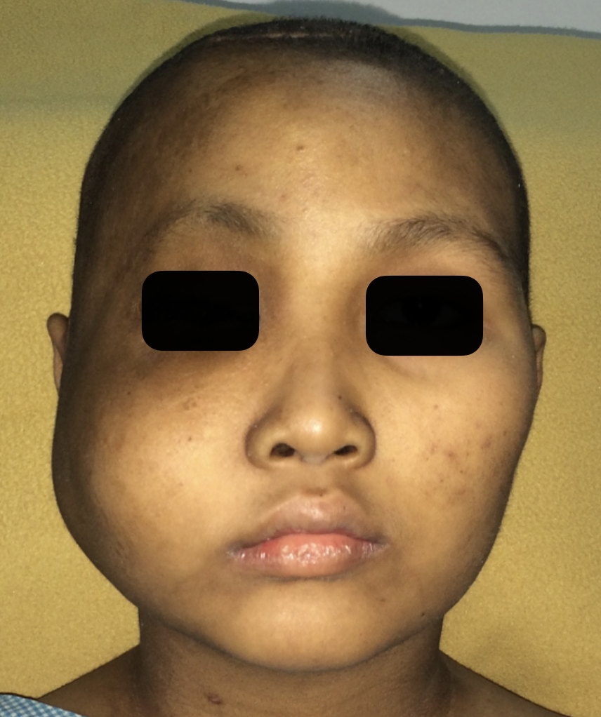
Fig.1 12 years old girl with osteogenic sarcoma. Presented with a large swelling in the region of righ cheek, eye and temple. She has lost the vision completely in the right eye.
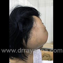
Fig.2 Right lateral view showing the extent of swelling
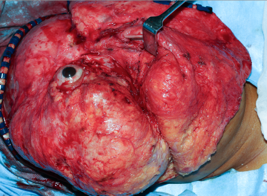
Fig.5 Skin flap is raised to expose the entire tumour and orbit.
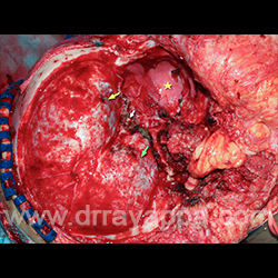
Fig.6 After complete excision of tumour involving skull & its base, maxilla and ethmoid.
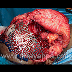
Fig.7 Defect in the skull was reconstructed using pre-shaped titanium mesh. Reconstruction was done using fibula osteocutaneous microvascular flap to regain the facial contour. Skin paddle was used to line the nasal cavity.
Fig.8 Clinical photograph at the time of discharge.
The Heart Of Clinic
Dr. C. Rayappa MBBS, DLO, FRCS(Edin)
SENIOR CONSULTANT
+91 44 3315 1105
Dr. C. Rayappa graduated from Madras Medical College, Chennai, India in 1982. He completed his post graduation in Otolaryngology (ENT)