JNA + Intracranial Extension Endoscopic Surgery + VIDEO
JNA + Intracranial Extension Endoscopic Surgery + VIDEO
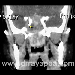
Fig.1 Coronal CT shows tumour filling the sphenoid and nasopharynx. Yellow arrow points at the defect in the lateral wall of sphenoid through which tumour entered the middle cranial fossa.
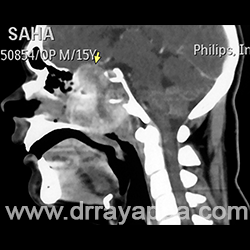
Fig.2 Sagital CT shows tumour filling posterior part of nasal cavity, nasopharynx, sphenoid and extending intracranially by eroding the bony roof of ethmoid and sphenoid (yellow arrow).
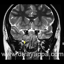
Fig.3 Coronal MRI shows the tumour in sphenoid extending through the defect in the lateral wall of sphenoid (yellow arrow) into middle cranial fossa.
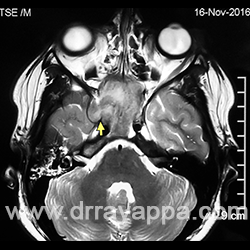
Fig.4 Axial MRI shows the tumour in sphenoid extending through the defect in the lateral wall of sphenoid (yellow arrow) into middle cranial fossa.
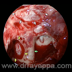
Fig.5 Picture of sphenoid sinus after resection of tumour. Green arrow – paraclival ICA. Yellow arrows point at the defect in the bony lateral wall of sphenoid. Cavernous sinus is seen through it. White arrows – defect in sphenoid roof.
Intracranial Extension Endoscopic Surgery
The Heart Of Clinic
Dr. C. Rayappa MBBS, DLO, FRCS(Edin)
SENIOR CONSULTANT
+91 44 3315 1105
Dr. C. Rayappa graduated from Madras Medical College, Chennai, India in 1982. He completed his post graduation in Otolaryngology (ENT)