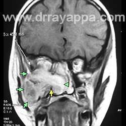
Fig.1 Coronal MRI shows tumour in the sphenoid sinus and parasellar region (green arrow). Yellow arrow – shows intact lateral wall of sphenoid.
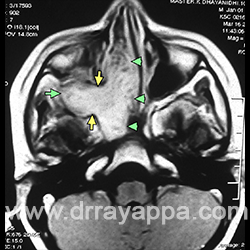
Fig.2 Coronal MRI shows tumour in the nose (green arrowhead) and sphenoid extending through the pterygomaxillary fissure (yellow arrow) and filling the entire infratemporal fossa (green arrowheads).
Fig.3 Axial MRI shows tumour in the nasal cavity (green arrowheads) extending through the pterygomaxillary fissure (yellow arrows) into infratemporal fossa (green arrow).
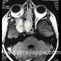
Fig.4 Axial MRI shows tumour in the sphenoid (arrowheads) and parasellar tumour (green arrow) with intact lateral sphenoid wall (yellow arrow).
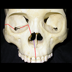
Fig.5 Picture shows osteotomy sites in maxilla. Osteotomy is done without stripping the soft tissues from which it receives its blood supply.
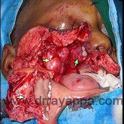
Fig.6 Intra-op picture after maxillary swing. Tumour in the nasal cavity (green arrow) and infratemporal fossa (yellow arrow) are exposed. White arrow – translocated maxilla.
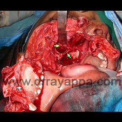
Fig.7 Intra-op picture after complete excision of tumour. Green arrow – lateral wall of sphenoid sinus. Retracting the middle fossa dura (yellow arrow) exposes the parasellar region that was occupied by the tumour.
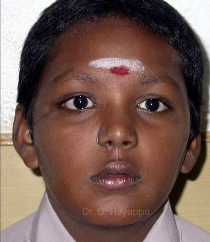
Fig.8 Post-op picture.
The Heart Of Clinic
Dr. C. Rayappa MBBS, DLO, FRCS(Edin)
SENIOR CONSULTANT
+91 44 3315 1105
Dr. C. Rayappa graduated from Madras Medical College, Chennai, India in 1982. He completed his post graduation in Otolaryngology (ENT)