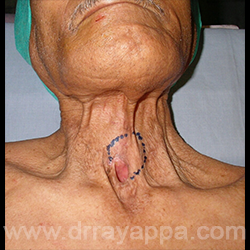
Fig.1 Seven years post laryngectomy and radiotherapy. Site of recurrence is marked.
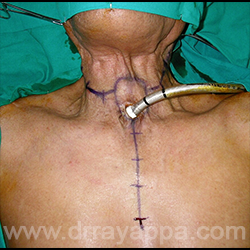
Fig.2 Skin incision is marked.
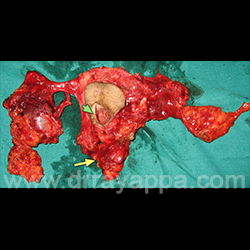
Fig.3 Operated specimen shows Bilateral neck dissection, superior mediastinal dissection (yellow arrow), trachea and the skin around it 9green arrow), pharynx and cervical oesophagus.
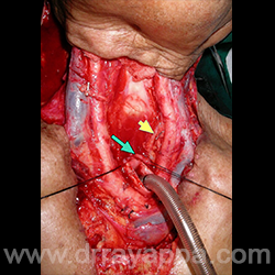
Fig.4 Picture shows vessels in the neck and superior mediastinum, thoracic duct (yellow arrow), cut end of oesophagus ( green arrow).
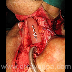
Fig.5 Jejunum microvascular flap is used to reconstruct the pharynx.
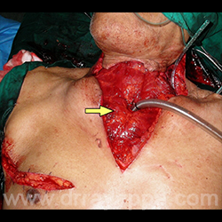
Fig.6 Pectoralis major muscle (yellow arrow)is rotated to cover the blood vessels and to create the tracheostoma.
The Heart Of Clinic
Dr. C. Rayappa MBBS, DLO, FRCS(Edin)
SENIOR CONSULTANT
+91 44 3315 1105
Dr. C. Rayappa graduated from Madras Medical College, Chennai, India in 1982. He completed his post graduation in Otolaryngology (ENT)