Mandibulectomy - Central Segment - Fibula Flap - Dental Implant
Mandibulectomy - Central Segment - Fibula Flap - Dental Implant
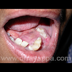
Fig.1 Tumour (ameloblastoma) involving most of the mandible 9molar to molar).
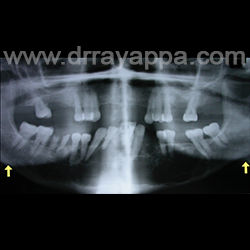
Fig.2 Panorex orthopantogram shows destruction of mandible from angle to angle (yellow arrows).
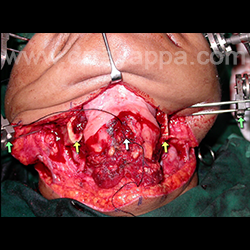
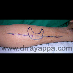
Fig.3 Mandible is resected from angle to angle. Yellow arrows – cut ends of mandible. Ramus of mandible were held in their original position with the external fixator (green arrows)for plating). White arrow – genioglossus muscle.
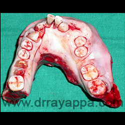
Fig.4 Specimen of the resected mandible from angle to angle.
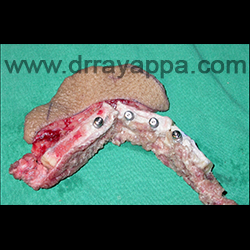
Fig.5 Fibula bone with the overlying skin is taken from the leg along with the vascular pedicle.
Fig.6 Fibula bone is cut into many pieces (without compromising vascularity) to create the natural shape of the mandible. Osseointegrated dental implants are fixed on the fibula bone.
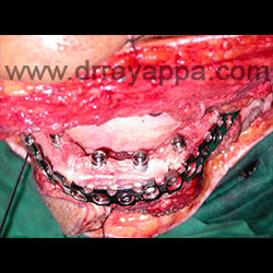
Fig.7 Shaped titanium reconstruction plates are screwed to the remaining ramus of mandible.Fibula bone with dental implants were screwed to the reconstrucion plate.
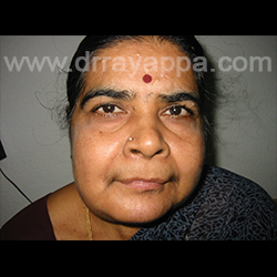
Fig.8 Good facial contour and symmetry.
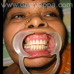
Fig.9 Denture is fixed on to the osseointegrated dental implants.
The Heart Of Clinic
Dr. C. Rayappa MBBS, DLO, FRCS(Edin)
SENIOR CONSULTANT
+91 44 3315 1105
Dr. C. Rayappa graduated from Madras Medical College, Chennai, India in 1982. He completed his post graduation in Otolaryngology (ENT)