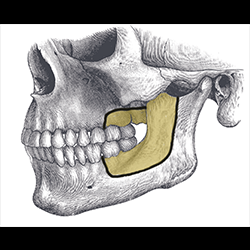
Fig.1 Rim resection of mandible. For tumours involving lower gingivobuccal sulcus and gingivam without bony erosion.
Fig.2 After raising lower cheek flap, tumour in the left buccal mucosa was excised with rim resection of mandible.
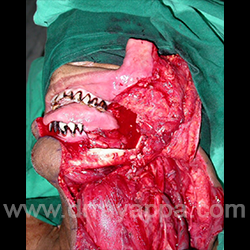
Fig.3 Excised specimen. white arrow – mandible. Green arrow – masseter muscle. Yellow arrow – buccal fat pad.
Fig.4 Eight yrs post-op picture. Good facial contour and symmetry.
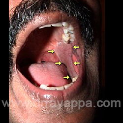
Fig.5 Post-op picture shows good mouty opening. Defect in the biccal mucosa was reconstructed with lateral arm microvascular flap (Yellow arrows).
Rim Resection without Lip Split
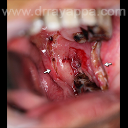
Fig.1 Ulcer in retromolar area (white arrows).
Fig.2 Mucosal cuts are made transorally 1 cm around the ulcer. Gingivam was incised between first and second molar. Anteriorly, the incision was extended along lower gingivobuccal sulcus. Soft tissues were elevated off the body of mandible.
Fig.3 Rim resection of mandible done which includes last 2 molars and anterior half of ramus (coloured area). Upper alveolus bearing the last 2 molars was resected (coloured area).
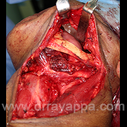
Fig.4 Lower cheek flap raised without splitting the lower lip is retracted shows intraoral structures after resection of tumour.
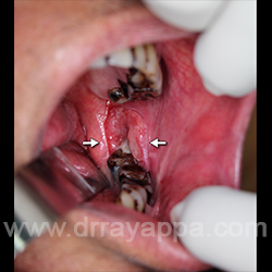
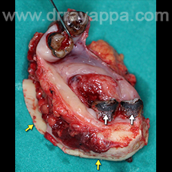
Fig.5 Resected specimen showing the mandible (yellow arrows), Lower molars (white arrows), tumour and upper molars (retracted with skin hook).
The Heart Of Clinic
Dr. C. Rayappa MBBS, DLO, FRCS(Edin)
SENIOR CONSULTANT
+91 44 3315 1105
Dr. C. Rayappa graduated from Madras Medical College, Chennai, India in 1982. He completed his post graduation in Otolaryngology (ENT)