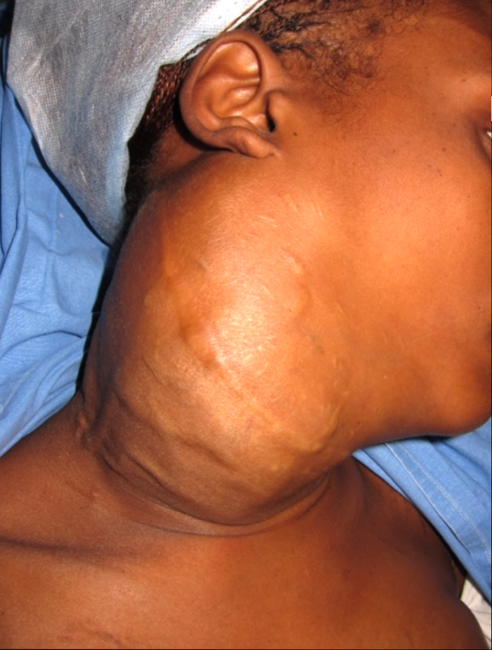
Fig.1 Large neck mass. Inferiorly extends up to the clavicle. Superiorly extends up to the ear lifting the lobule of the ear.
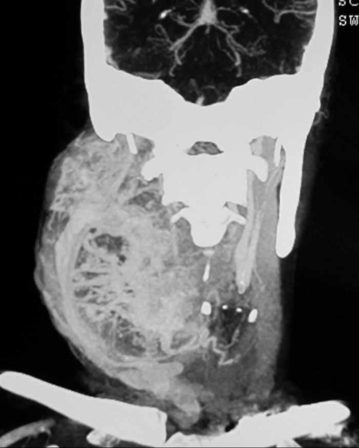
Fig.2 CT angio shows a hypervascular mass extending from clavicle to the skull base surrounding the CCA, ECA & ICA. Temporary baloon occlusion test revealed good cross circulation through circle of willis. Therefore, ICA was occluded permanently.
Fig.3 Coronal MRI shows the extent of tumour. Green arrow – Condyle
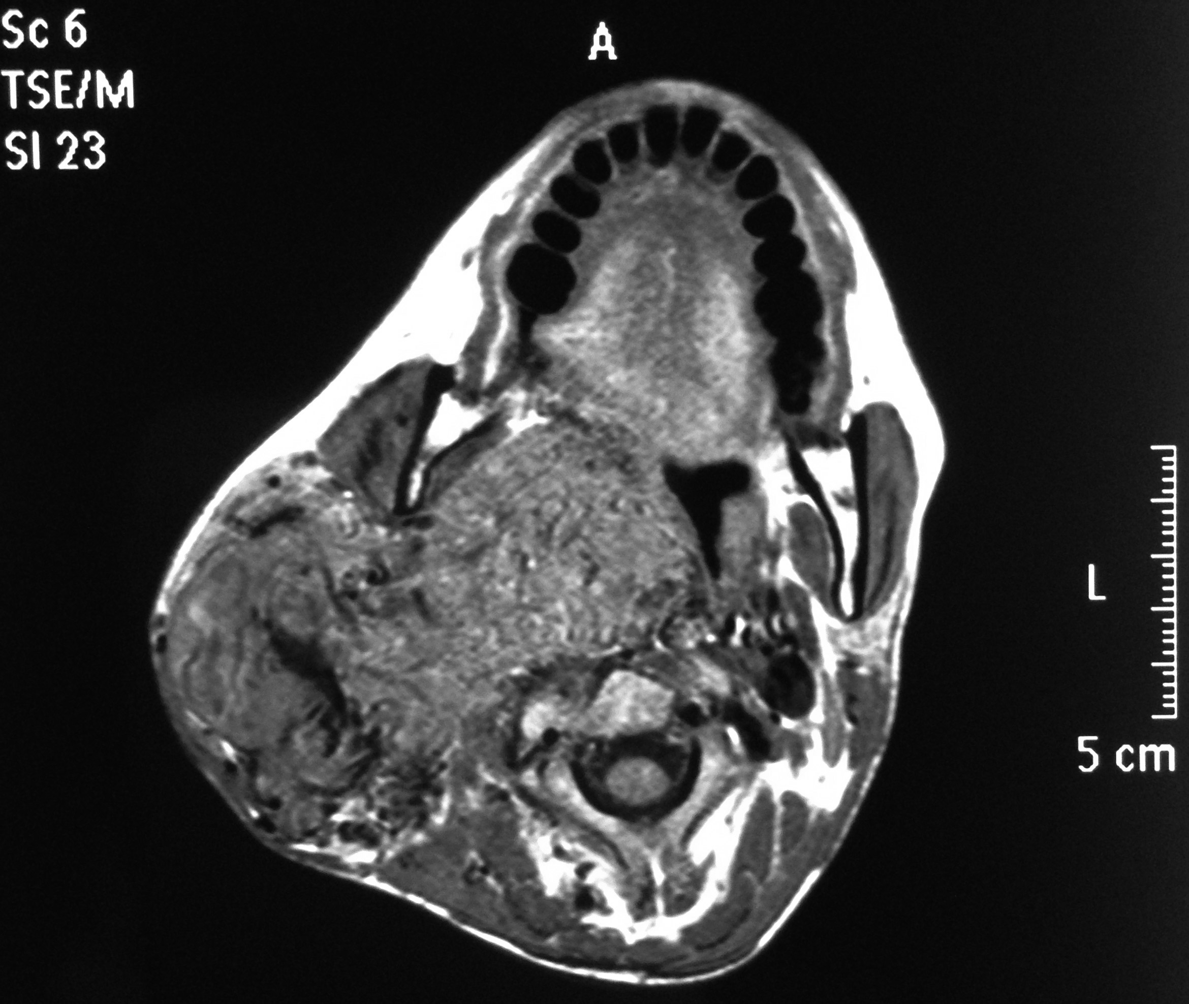
Fig.4 Axial MRI shows the tumour extent and the narrowed oropharyngeal airway.
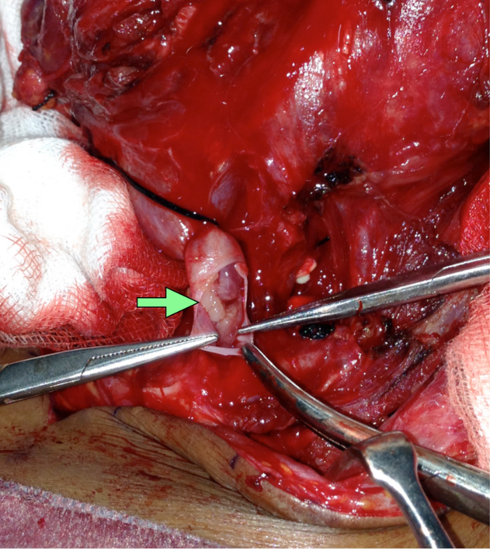
Fig.5 Mandibulotomy was done to expose the tumour reaching the skull base. Yellow arrow – site of mandibulotomy. Green arrow – internal jugular vein.
Fig.6 Tumour within the lumen of IJV
Fig.7 Tumour in the IJV was found extending into the subclavian vein and was pulled out (yellow arrows). Green arrow – subclavian vein.
Fig.8 Excised tumour. Forceps is holding the lower cut end of IJV. Green arrow – tumour pulled out of lower part of IJV. Yellow arrow – tumour pulled out from subclavian vein.
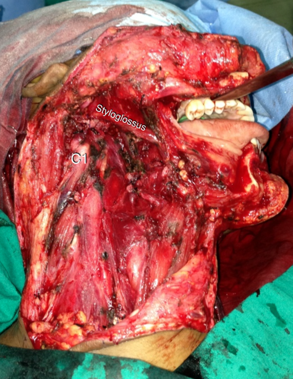
Fig.9 After complete excision of tumour along with CCA, ECA, ICA, IJV and lower cranial nerves
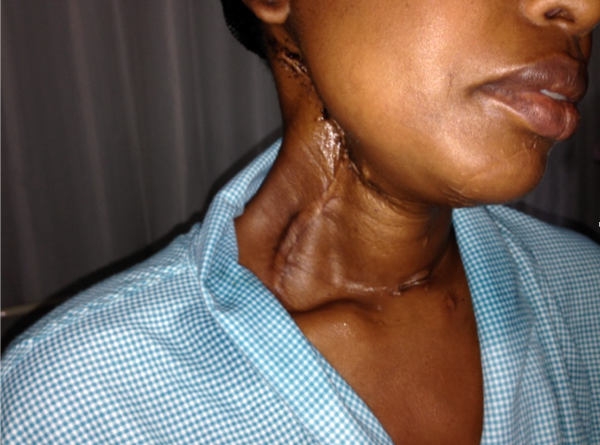
Fig.10 Her swallowing and speech improved after 3 weeks. This picture was taken 6 months later when she came for follow up.
The Heart Of Clinic
Dr. C. Rayappa MBBS, DLO, FRCS(Edin)
SENIOR CONSULTANT
+91 44 3315 1105
Dr. C. Rayappa graduated from Madras Medical College, Chennai, India in 1982. He completed his post graduation in Otolaryngology (ENT)