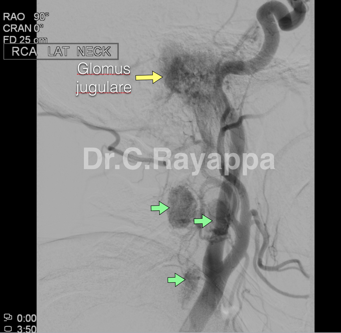
Fig. 1 Carotid angiogram was done for pre-op embolization. In addition to glomus jugulare, it revealed three hypervascular areas in the neck. These nodes were found to have glomus tissue.
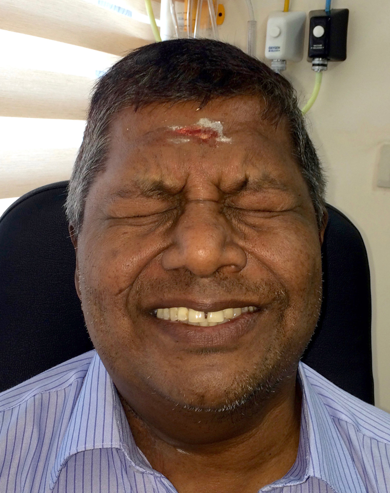
Fig.2 Patient underwent ITF type-A approach as well as neck dissection. Minimal facial paresis + immediately after surgery.
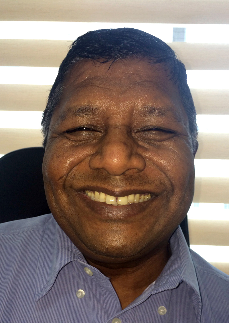
Fig.3 Full recovery of facial movements 6 months after surgery.
Fig.4 Full recovery of facial movements 6 months after surgery.
The Heart Of Clinic
Dr. C. Rayappa MBBS, DLO, FRCS(Edin)
SENIOR CONSULTANT
+91 44 3315 1105
Dr. C. Rayappa graduated from Madras Medical College, Chennai, India in 1982. He completed his post graduation in Otolaryngology (ENT)