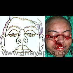
Fig.2 Incisions as marked in the diagram. Between medial canthus and lateral canthus, the incision goes through the inferior fornix of conjunctiva (interrupted lines).
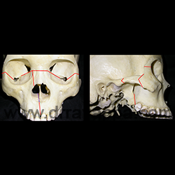
Fig.3 Osteotomies were made as in the picture without stripping the soft tissues from maxilla.
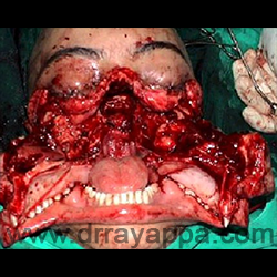
Fig.4 Tumour is removed after translocating nose and both maxillae.
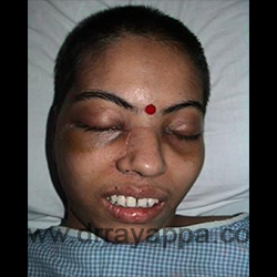
Fig.5 2 weeks post-op. Patient has regained the facial contour, good occlusion and taking food by mouth. Scar is barely noticeble.
Midfacial split VIDEO
The Heart Of Clinic
Dr. C. Rayappa MBBS, DLO, FRCS(Edin)
SENIOR CONSULTANT
+91 44 3315 1105
Dr. C. Rayappa graduated from Madras Medical College, Chennai, India in 1982. He completed his post graduation in Otolaryngology (ENT)