Fig.1 Adenocarcinoma of nasopharynx & ITF. There is a fat plane between the tumour and ICA (yellow arrow).
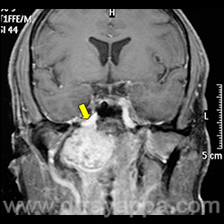
Fig.2 MRI shows the proximity of the tumour to the ICA (yellow arrow).
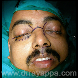
Fig.3 Incision as marked. From medial to lateral canthus, the incision extends through the inferior fornix of conjunctiva (interrupted line) to preserve the facial nerve to orbicularis oculi & to avoid the scar.
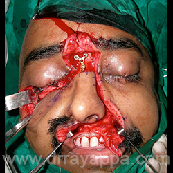
Fig.4 Soft tissues are elevated minimally to expose the site of osteotomy. Excessive dissection may devitalize the bone. Osteotomy sites are marked and preplating done.
Fig.5 Osteotomy sites are marked.
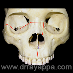
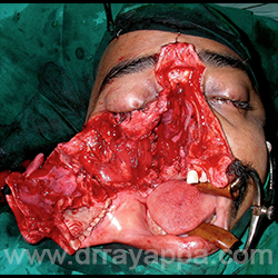
Fig.6 Nose and maxilla are rotated as a single unit with the soft tissues attached. Gives a wide exposure of nose, nasopharynx and ITF.
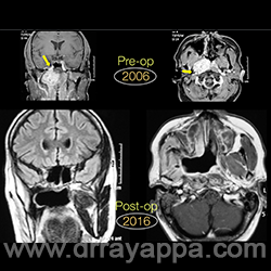
Fig.7 Pre-op and 10 yrs post-op scan. No evidence of recurrence.
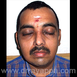
Fig.8 Good facial symmetry, good occlusion & mouth opening and facial nerve function. Inconspicuous scar.
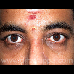
Fig.9 Closeup showing thin scar over the nose.
The Heart Of Clinic
Dr. C. Rayappa MBBS, DLO, FRCS(Edin)
SENIOR CONSULTANT
+91 44 3315 1105
Dr. C. Rayappa graduated from Madras Medical College, Chennai, India in 1982. He completed his post graduation in Otolaryngology (ENT)