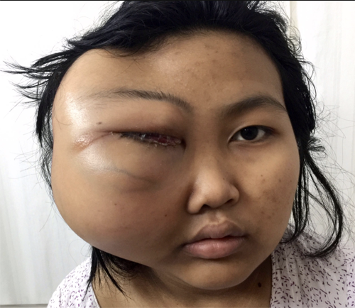
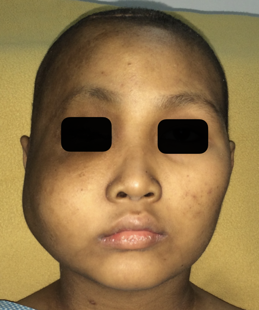
Fig.1. 12 years old girl with osteogenic sarcoma. Presented with a large swelling in the region of right cheek, eye, and temple. She has lost vision completely in the right eye.
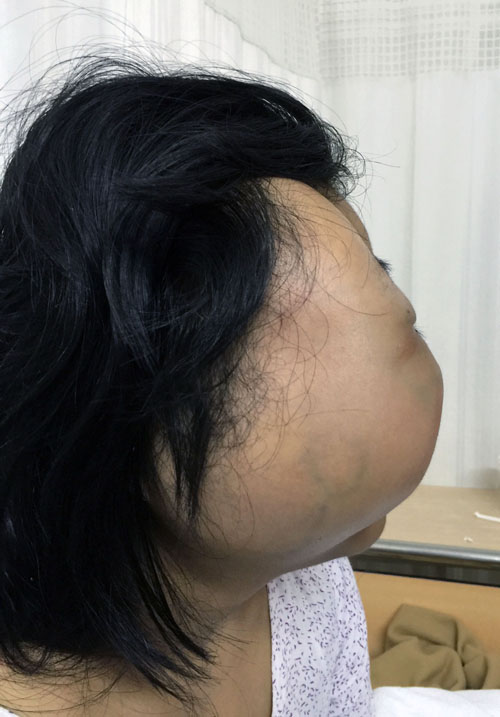
Fig.2. Right lateral view showing the extent of swelling
Fig.3. MRI shows tumour involving the temporal fossa (yellow arrow), zygoma & orbit (white arrow), and ethmoid sinus (green arrow)
Fig.4. MRI shows tumour involing temporal fossa (white arrow), cheek & infratemporal fossa (yellow arrow), nasal cavity, ethmoid and max
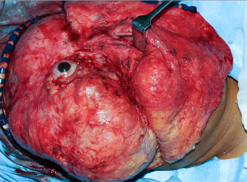
Fig.5. Skin flap is raised to expose the entire tumour and orbit
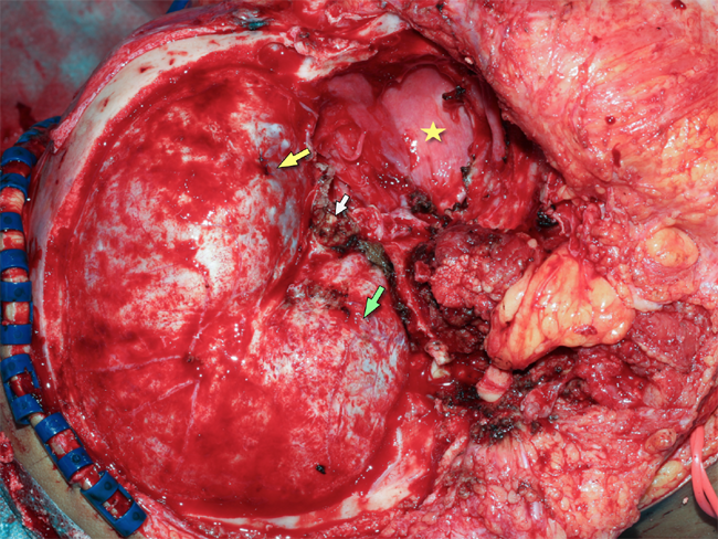
Fig.6. After complete excision of tumour involving the skull & its base, maxilla, and ethmoid
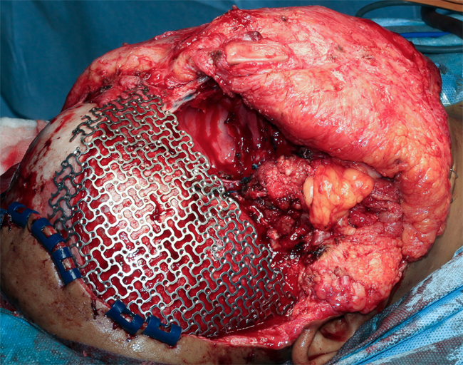
Fig.7. Defect in the skull was reconstructed using pre-shaped titanium mesh. Reconstruction was done using a fibula osteocutaneous microvascular flap to regain the facial contour. A skin paddle was used to line the nasal cavity.