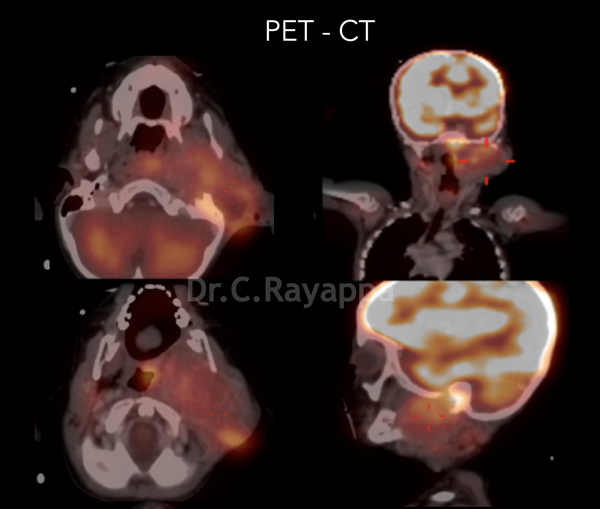
Fig.2 PET CT shows tumour extending into infratemporal fossa
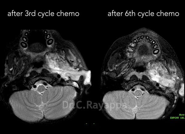
Fig.3 MRI shows minimal response to chemotherapy
Fig.4 Residual tumour after 6 cycles of chemotherapy
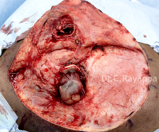
Fig.5 Skin flap is raised
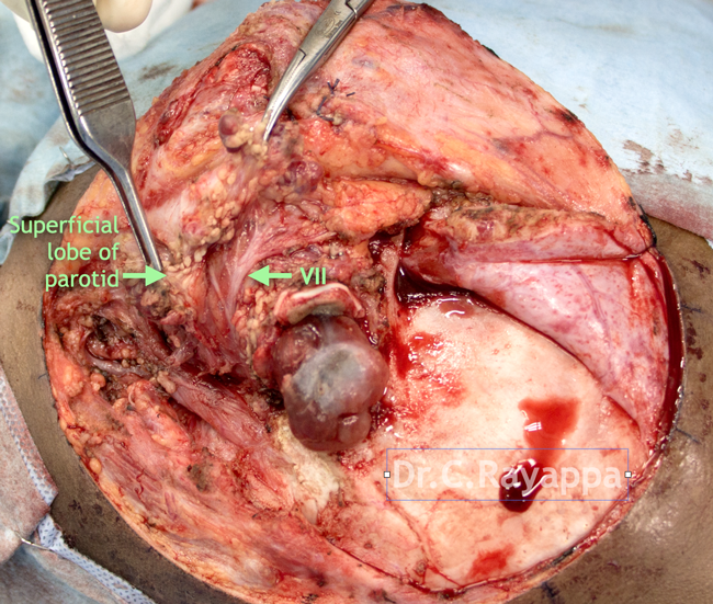
Fig.6 Superficial lobe of parotid was dissected off the facial nerve and its branches.
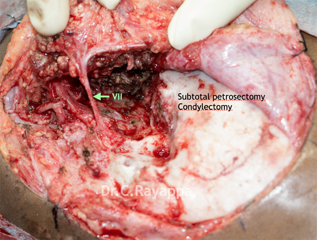
Fig.7 Subtotal petrosectomy had been done
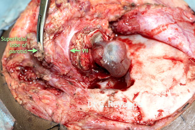
Fig.8 Condyle of the mandible and content of the infratemporal fossa had been resected along with the deep lobe of the parotid. The facial nerve and its branches were preserved
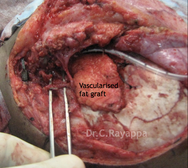
Fig.9 Defect was reconstructed using vascularised fat
Fig.9 Defect was reconstructed using vascularised fat
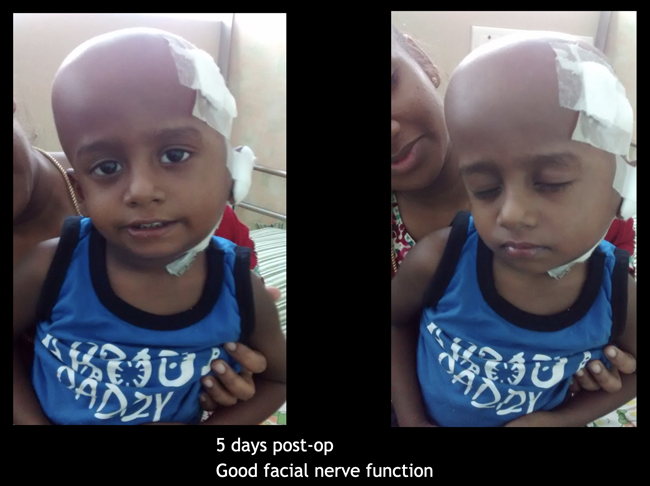
Fig.10 5 days after surgery. Good facial nerve function
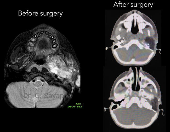
Fig.11 Scans done after surgery shows complete resection of tumour. The fat graft used appears black in the CT scan
The Heart Of Clinic
Dr. C. Rayappa MBBS, DLO, FRCS(Edin)
SENIOR CONSULTANT
+91 44 3315 1105
Dr. C. Rayappa graduated from Madras Medical College, Chennai, India in 1982. He completed his post graduation in Otolaryngology (ENT)