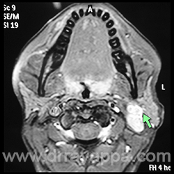
Fig.1 Axial MRI. Tumour involving the deep lobe of parotid gland. Absence of fat plane (green arrow) between the mass and the superficial lobe of parotid gland indicates it’s a deep lobe tumour.
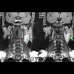
Fig.2 Coronal MRI. Tumour involving the deep lobe of parotid gland. Absence of fat plane (green arrow) between the mass and the superficial lobe of parotid gland indicates it’s a deep lobe tumour
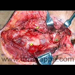
Fig.3 After identifying the facial nerve trunk (green arrow), superficial lobe of the parotid gland (yellow arrow) is dissected off the facial nerve.Facial nerve is seen upto the bifurcation. Yellow star – tumour.
Fig.4 Superficial lobe of the parotid gland (yellow arrow) is dissected off the lower division of facial nerve (green arrow) and marginal mandibular nerve (green arrow head).
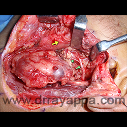
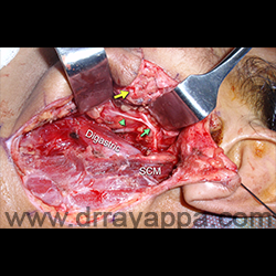
Fig.5 After resection of tumour. SCM – sternocledomastoid muscle. Digastric – posterior belly. Green arrow – lower division of facial nerve. Green arrowhead – marginal mandibular nerve. Superficial lobe (yellow sarrow) is laid back.
The Heart Of Clinic
Dr. C. Rayappa MBBS, DLO, FRCS(Edin)
SENIOR CONSULTANT
+91 44 3315 1105
Dr. C. Rayappa graduated from Madras Medical College, Chennai, India in 1982. He completed his post graduation in Otolaryngology (ENT)