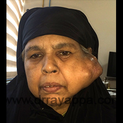
Fig.1 60+ yrs old patient has been having this large, hard and multi lobulated mass infront of her left ear. Gradually increased in size over the past 10 years. FNAC was reported as pleomorphic adenoma.
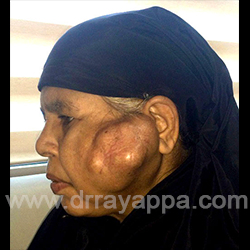
Fig.2 Tumour was involving entire parotid gland. Skin was stretched and thinned out. It was adherent to the tumour at certain areas. Facial nerve function was intact.
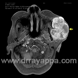
Fig.3 Axial MRI shows the extent of tumour. Skin is infiltrated at the summit of tumour (yellow arrow).
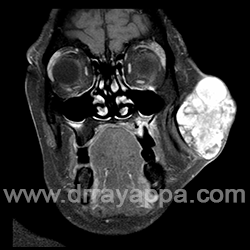
Fig.4 Coronal MRI.
Fig.5 Skin incision.Skin over the posterior half of tumour will be excised to bring the suture line as close to the ear as possible (white arrow).
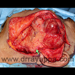
Fig.6 Skin flaps were elevated. Tumour is found involving the upper part of the parotid gland. Green arrow – uninvolved lower part of the parotid gland.
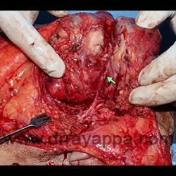
Fig.7 Buccal branch of the facial nerve (green arrow) was going through the tumour. Tumour was split to dissect out the buccal branch.
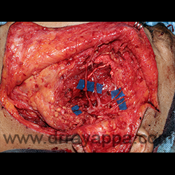
Fig.8 After removal of the tumour. Facial nerve trunk and all its branches were preserved (blue sheet placed under the nerves for contrast).
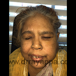
Fig.9 One week post surgery. Temporary mild facial paresis on left side.
The Heart Of Clinic
Dr. C. Rayappa MBBS, DLO, FRCS(Edin)
SENIOR CONSULTANT
+91 44 3315 1105
Dr. C. Rayappa graduated from Madras Medical College, Chennai, India in 1982. He completed his post graduation in Otolaryngology (ENT)