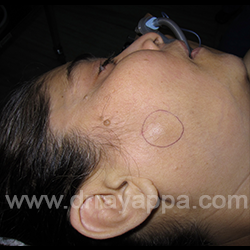
Fig.1 This patient presented with a 2cm, non-tender, mobile nodule in her right cheek (circle). FNAC was reported as mucoepidermpoid carcinoma.
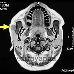
Fig.2 Axial MRI shows the tumour (yellow arrow) lying on masseter muscle anterior to parotid gland.
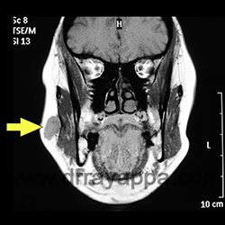
Fig.3 Coronal MRI shows the tumour (yellow arrow) lying over the masseter muscle. Parotid salivary gland is not visible in this picture.
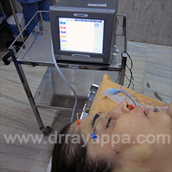
Fig.4 Nerve integrity monitor was used to help in localizing the facial nerve branches. Electrodes inserted into different facial muscle groups (orange, blue, red & green colours).
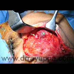
Fig.5 Skin flap raised anterior to the tumour (green star).Blunt dissection was done to identify the buccal branch of facial nerve and parotid duct (yellow arrow).
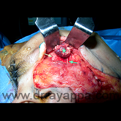
Fig.6 Picture shows the completely dissected out tumour (green star), parotid duct (yellow arrow) and buccal branch of facial nerve (green arrow).
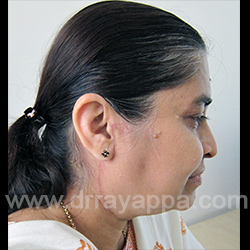
Fig.7 Post-op picture shows good facial nerve function.
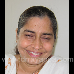
Fig.8 Post-op photograph. Barely noticeble scar.
The Heart Of Clinic
Dr. C. Rayappa MBBS, DLO, FRCS(Edin)
SENIOR CONSULTANT
+91 44 3315 1105
Dr. C. Rayappa graduated from Madras Medical College, Chennai, India in 1982. He completed his post graduation in Otolaryngology (ENT)