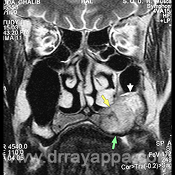
Fig.1 MRI shows tumour involving lower part of maxillary antrum (white arrow), nasal floor (yellow arrow) and palate & alveolus (green arrow).
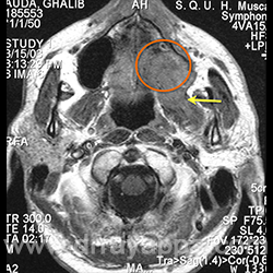
Fig.2 MRI shows tumour involving the maxillary antrum (orange circle) and infiltrating pterygoid muscle (yellow arrow).
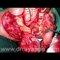
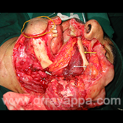
Fig.3 Upper and lower cheekflaps were raised exposing mandible, maxilla and zygomatic arch. IOF – infraorbital foramen, ZA – zygomatic arch, CP – coronoid process, TMJ – temperomandibular joint, Yellow arrow – osteotomy sites.
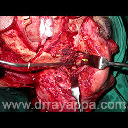
Fig.4 Mandible was divided at the angle (white arrow). The cut zygomatic arch was retracted and the temporalis muscle was divided at the temporal fossa. This exposes the greater wing of sphenoid in temporal fossa (yellow arrow).
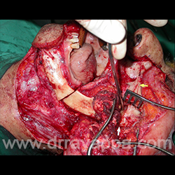
Fig.5 Site of osteotomy seen in the anterior wall of maxilla (yellow arrow). Temperomandibular joint is disarticulated (white arrow) and the infratemporal fossa contents are dissected subperiosteally from greater wing of sphenoid.
Fig.6 Infrastructure maxillectomy and ITF clearance had been done. Yelloe arrow – greater wing of sphenoid. White arrow points at foramen ovale.
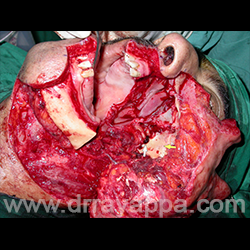
Fig.7 Microvascular flap (white arrow) was used to cover the defect in palate and fill the dead space in ITF. Bone graft was used to provide facial contour.
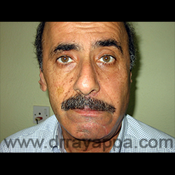
Fig.8 Post- op picture showing good facial contour and symmetry.
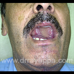
Fig.9 Picture shows the microvascular flap covering the defect in the palate.
The Heart Of Clinic
Dr. C. Rayappa MBBS, DLO, FRCS(Edin)
SENIOR CONSULTANT
+91 44 3315 1105
Dr. C. Rayappa graduated from Madras Medical College, Chennai, India in 1982. He completed his post graduation in Otolaryngology (ENT)