drrayappa.com

Total Maxillectomy

Total Maxillectomy

Fig.1 The incision used by the author is marked. It gives sufficient exposure for maxillectomy. The lateral extension along the lower eyelid used traditionally gives an ugly scar and is not required.
Fig.2 Osteotomy sites for total maxillectomy are marked.
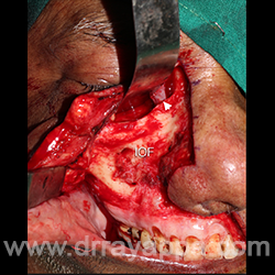
Fig.3 Skin flap is raised exposing anterior wall of maxilla, zygoma and frontal process of maxila. Malleable retracting the orbital content exposing the orbital floor and inferior orbital fissure. IOF – infraorbital foramen. Arrow head – Lacrimal sac.
Fig.4 Picture shows maxillectomy defect. Orbital floor is reconstructed with titanium mesh and is covered with vascularised soft tissue.
Fig.5 Total maxillectomy defect had been reconstructed with fibula microvascular flap (different patient). Good facial contour, less visible scar.

The Heart Of Clinic

Dr. Rayappa

Dr. C. Rayappa MBBS, DLO, FRCS(Edin)

SENIOR CONSULTANT

+91 44 3315 1105

Dr. C. Rayappa graduated from Madras Medical College, Chennai, India in 1982. He completed his post graduation in Otolaryngology (ENT)

The Heart Of Clinic

Dr. Rayappa

Dr. C. Rayappa MBBS, DLO, FRCS(Edin)

SENIOR CONSULTANT

+91 44 3315 1105

Dr. C. Rayappa graduated from Madras Medical College, Chennai, India in 1982. He completed his post graduation in Otolaryngology (ENT)